
Spontaneous biliary perforations in children are rare and most of the reported cases are from perforation of Choledochal cyst. Pseudocyst formation simulating a choledochal cyst from spontaneous perforation of the apparently normal biliary tree is unusual.
A four-month-old female child born to nonconsanguineous parents was brought to the hospital with a history of progressive abdominal distension, passing white-colored stool, and high colored urine of one-week duration.
The child had surgery for a bilateral inguinal hernia at two months of age. Clinical examination showed that the child was alert and active with pallor and icterus. The abdomen was grossly distended, with dilated veins over the anterior abdominal wall. Free fluid was present in the abdomen.
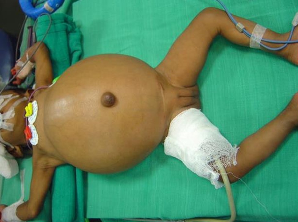
Neonate with grossly distended abdomen and gross ascites
Blood counts were elevated, and the liver function tests showed elevated bilirubin with direct conjugated hyperbilirubinemia. The total bilirubin was 6.2mg %, direct bilirubin 5.0mg%, total
protein 5.1gm%.
Pre-operative Ultrasound abdomen showed a thin gall bladder, large serpiginous cystic mass under the surface of liver extending from porta hepatis to the tip of the left lobe of the liver.
Management
Through a right subcostal incision the abdomen was opened and nearly 200 ccs of bile-stained fluid were sucked out. There were adhesions between the liver and the colon. The gall bladder was collapsed. A thick-walled cyst was seen on the right side of the porta hepatis. Aspiration drew bile and an operative cholangiogram was done.
It showed a normal intrahepatic biliary tree with the dilated upper portion of the common bile duct. The rest of the common bile duct was replaced by a large transversely situated cyst. The distal common bile duct was not seen and there was no flow of dye into the duodenum. The cyst was drained by an 8F Foley catheter. Liver biopsy was done.
The histology of the liver was normal. The bile discharge from the drain continued for nearly 15 days and the motion remained white-colored. The serum bilirubin returned to normal. The child was re-explored through the original incision. There were dense adhesions between the liver and the first portion of the duodenum. The original cyst was not seen. As the adhesion was released there was an incidental small perforation in the first part of the duodenum. A dense stricture was seen extending from the common hepatic duct to the junction with the cystic duct The common bile duct distal to cystic duct was normal. The structured segment with the gall bladder was removed. Cholangiogram done through the distal common bile duct showed the free flow of the dye into the duodenum.
The two cut ends of the common bile duct were anastomosed over a 5 French feeding tube with a side hole. It was brought through the incidental perforation in the first part of the duodenum and drained externally. The complete disappearance of the cyst during the second surgery indicated the cyst which was seen during the first exploration was a pseudocyst simulating Choledochal cyst. On the fifteenth day after the second surgery cholangiogram was done through the feeding tube draining the common bile duct showed the free flow of bile into the duodenum and normal proximal ducts with the minimal periductal leak. The drainage tube was removed and the child made a good recovery. Follow up after two years showed normal liver function tests with normal growth and development.
Get Care
Getting an appointment with Dr. Ajay Kumar Verma is easy. Schedule using any of these convenient options.